Surgical treatment
The repair of a wall defect is built on three choices: the access route (open, laparoscopic, robotic or endoscopic), the type of mesh and the plane in which it is placed. This page describes the main techniques, from cornerstones such as Rives-Stoppa to the component separations.
This page contains images of surgical procedures. Viewing is not advised for those who are easily affected.
Which techniques are used today for the abdominal wall?
Operations to repair defects of the anterior abdominal wall can be performed by an open route or by a minimally invasive route.
Open surgery of the anterior abdominal wall is very heterogeneous. It ranges from procedures with small incisions to repair a tiny umbilical hernia without a mesh, to very complex operations with large incisions lasting several hours, to repair wall disasters. Repair of the wall with elimination of the defect can be achieved by direct suture or with the help of prosthetic materials that make the repair more solid.
Currently very few conditions do not involve the use of a mesh. While a small hernia of 1–1.5 cm can be repaired by direct suture, albeit with a high recurrence risk, the decision to use an open approach and whether or not to use a mesh is based on a careful assessment of the patient's age, history and risk factors.
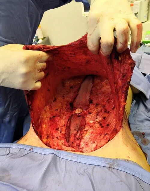
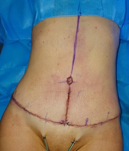
In recent years open surgery has given much ground to minimally invasive surgery. Setting aside wall disasters and emergency surgery, almost every type of operation on the anterior abdominal wall has by now been performed through a minimally invasive route.
The minimally invasive techniques are many: several have been developed in recent years, each with its own features. This has markedly raised the level and quality of this type of surgery. Most recently, robotic surgery.
Among the minimally invasive techniques used to repair the abdominal wall are: eTEP, THT, MISAR, TAPP, TESAR and SCOLA.
Beyond the acronyms, these techniques can be classified by the position of the instruments relative to the peritoneal cavity: laparoscopic, operating with instruments inside the cavity; endoscopic, working outside the cavity; endo-laparoscopic, a mix of the two; and robotic, using automated platforms. They can also be divided, more technically, by their approach to the muscular wall, which may be posterior (from behind or beneath the muscles) or anterior (from above or in front).
The posterior approach can be further categorised as intraperitoneal (from inside the abdominal cavity) and extraperitoneal (outside the cavity but behind the muscles). There are no definitive data on which technique is superior, and each has its pros and cons. The choice of technique today depends on the surgeon's preferences but should depend on the type of defect to treat and the result to achieve.
What is the posterior intraperitoneal approach?
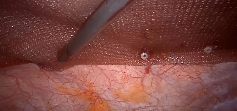
IPOM (Intra Peritoneal Onlay Mesh) is now considered an imprecise term; IPUM (Intraperitoneal Underlay Mesh) is preferred to better reflect the intraperitoneal placement of the mesh. The traditional IPOM technique uses a mesh to cover the defect but not truly repair it, which has generated debate about its effectiveness. It is not suitable, for example, for correcting diastasis, since it does not solve the muscular defect.
To address these limits, a variant called IPOM PLUS was introduced, which includes suturing the defect before placing the mesh in the peritoneal cavity.
A further evolution is the LIRA technique, published by an important Spanish group, a variation of IPOM PLUS. It involves incising the posterior rectus sheath at a certain distance from the defect and aims to reduce tension on the suture line, making the whole procedure safer.
What is the posterior endo-laparoscopic approach?
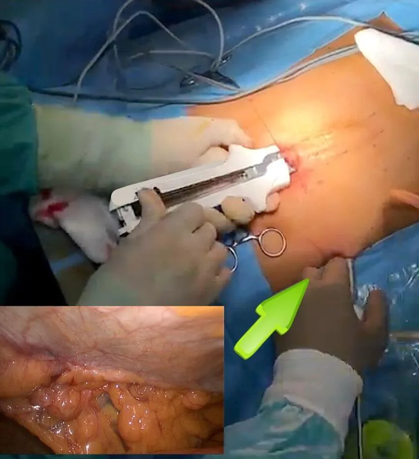
THT is a technique devised by Dr. Alessandro Carrara, inspired by Dr. Thiago Costa's stapler technique. Using a single umbilical access and a camera for vision, Dr. Carrara simplified the procedure, making it more reproducible. The technique focuses on the simultaneous incision and suture of the muscles and fasciae with a mechanical stapler, completing the operation with a retromuscular mesh.
The MISAR technique, devised by Dr. Gabriele Manetti, follows an approach similar to Costa's. It involves three suprapubic incisions with intraperitoneal access and a mechanical suture to bring the muscles together, repair the defect and create space for the mesh.
What is the posterior extraperitoneal endoscopic approach?
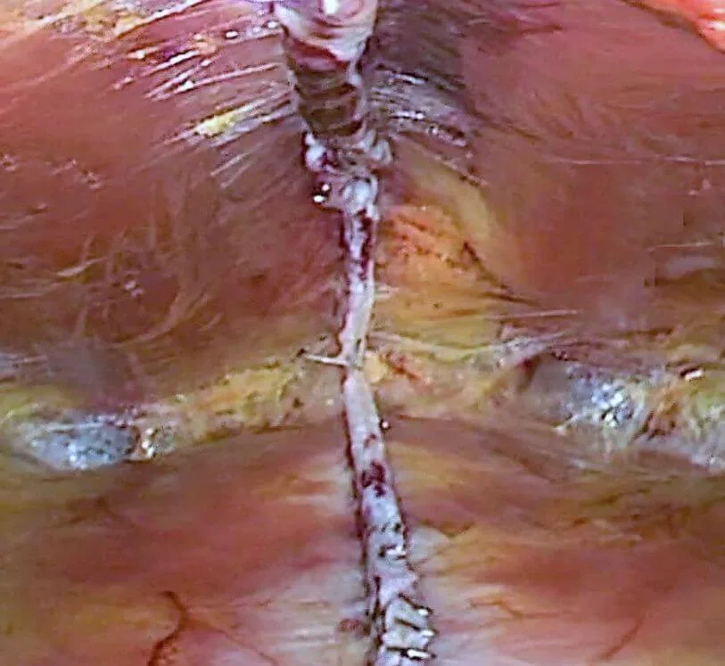
The posterior extraperitoneal approach (eTEP, enhanced-view Totally Extra Peritoneal) is currently the most talked-about technique in minimally invasive surgery. It does not involve, at least initially, the abdominal cavity, but rather a surgically created retromuscular area. It is used both for midline defects and for inguinal and femoral ones. Since its introduction in the early 1990s the technique has seen significant evolution, in particular in 2012 with the contribution of Professor Jorge Daes. Together with Daes, Belyansky, Radu, Ramana, Novitsky and other leading international experts developed it further in March 2018, making it possible to repair various types of hernia, not only inguinal.
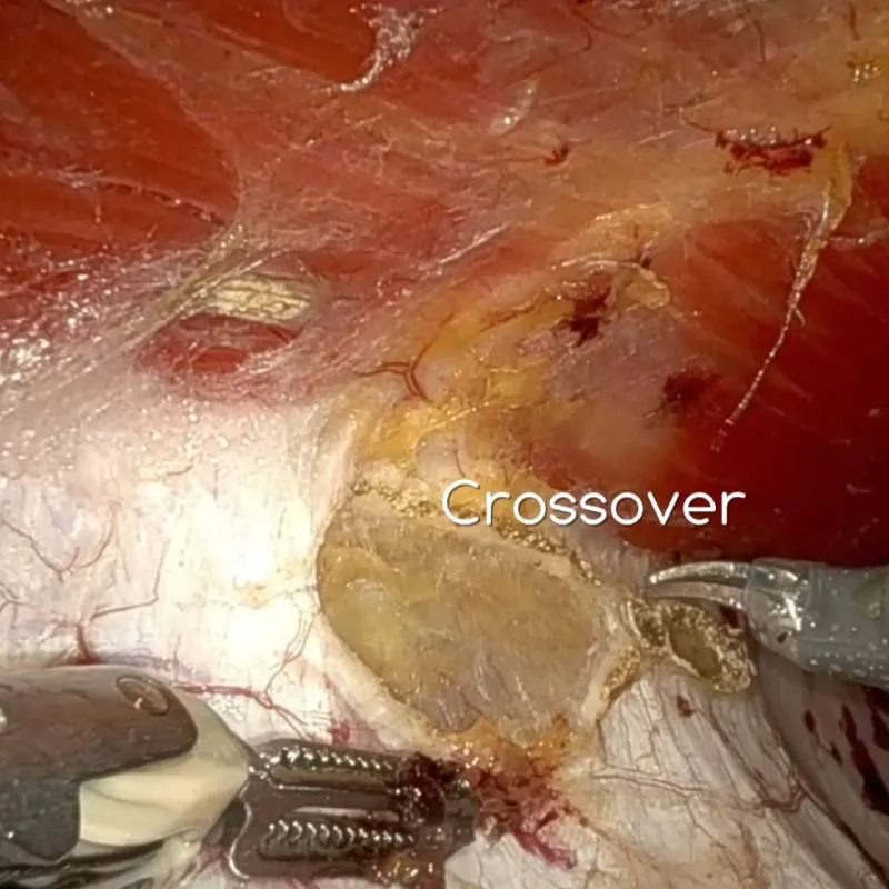
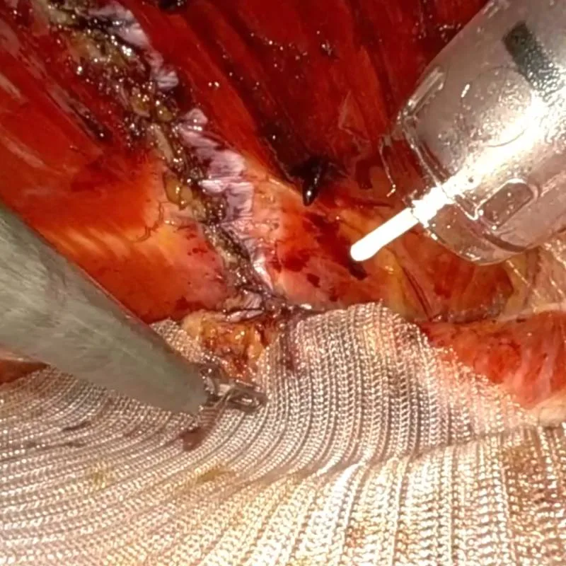
The procedure begins with access to the retromuscular area using a camera and other instruments. A unified working space is created behind the muscles, crossing from one side to the other of the retrorectus zone with a technique called "crossover". The defect is then repaired, from both the posterior and anterior sides, using various sutures. Finally a mesh is placed, usually fixed with glue, before the instruments are removed and the operation completed. In most secondary repairs, that is incisional hernias, it is not possible to complete the procedure remaining entirely extraperitoneal, which in some cases can entail risks; the procedure must therefore be performed by experienced hands.
The evolution of this technique today involves the robotic platform (r-eTEP). The use of robotic platforms has greatly simplified the procedure, which is otherwise complicated by the need to manoeuvre the instruments "in reverse".
What is the anterior approach?
The anterior approach to minimally invasive surgery of the anterior abdominal wall was essentially introduced in 2014 by Bellido and Guadalajara. These Spanish surgeons used small incisions and endoscopic instruments to dissect the tissue above the muscular fascia, creating a safe and ample working space. Their study presented 21 patients treated with a simple continuous suture to repair diastasis and possible hernias.
What is the onlay, supra-muscular mesh?
This early experience inspired other techniques, including REPA (Reparación Endoscópica Pre-Aponeurótica) by Derlin Muas and SCOLA (Sub-Cutaneous Onlay Laparoscopic Approach) by Flavio Malcher, both based on an extended prefascial dissection and the addition of a mesh above the muscles (onlay). Both have received scientific recognition and are considered essentially identical.
What is the sublay, retromuscular mesh?
TESAR (Totally Endoscopic Sublay Anterior Repair), presented by our group in 2018 and published in 2019, is the anterior endoscopic technique with a retromuscular sublay mesh developed for midline defects. The full description of the technique, the published scientific data and the results are the canonical source on federicofiori.com: the TESAR technique.
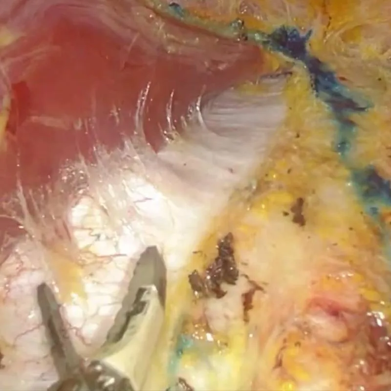
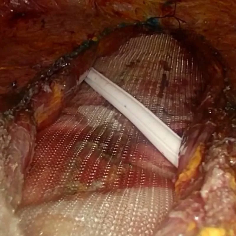
To minimise the risk of seroma, a subcutaneous and fascial suture is performed to reduce the dead space, and a prefascial drain is placed. After the trocars are removed and the incisions sutured, a compressive dressing is applied, which should be worn for at least 20–30 days.
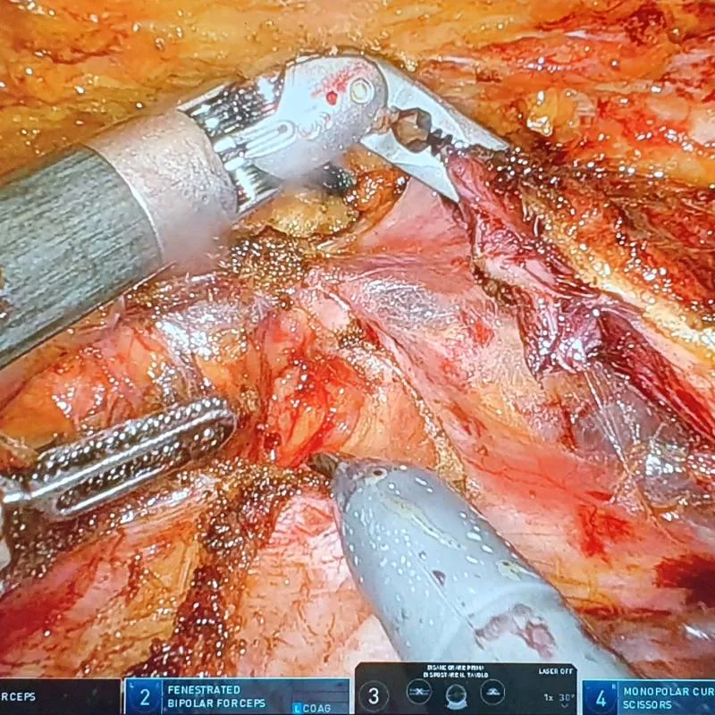
What changes with robotic surgery?
Robotics is in fact a form of minimally invasive approach; today it is a real option and is strongly indicated in abdominal wall repair, with important advantages for the patient and the surgeon in terms of precision, safety and ergonomics.
The main advantages of a robotic platform are:
Precision and control
- Robotic systems offer markedly improved precision and control compared with traditional techniques.
- Robotic instruments can reach difficult areas and operate with a precision beyond the capacity of the human hand, reducing the risk of damage to surrounding tissues.
Improved visualisation
- Robotic surgery offers high-definition 3D visualisation of the surgical field, giving a clear and magnified view.
- This helps identify and protect critical structures during the repair of the wall.
Reduced surgeon fatigue
- Long procedures can be physically demanding for a surgeon.
- Robotic systems let surgeons work in a more comfortable position, reducing fatigue and potentially improving precision during prolonged operations.
Minimally invasive access
- Robotic surgery allows minimally invasive access, potentially reducing postoperative pain and recovery times.
Application in complex cases
- In some complex cases of abdominal wall repair, robotic surgery can provide solutions that would otherwise be very difficult or impossible with conventional techniques.
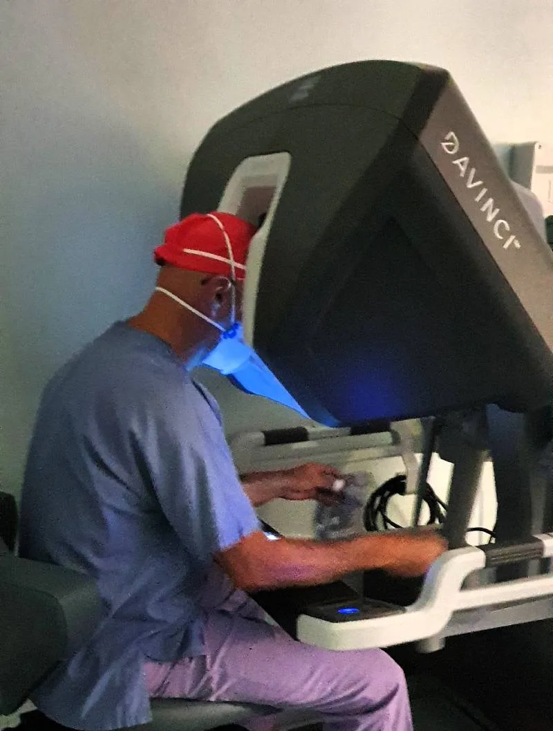
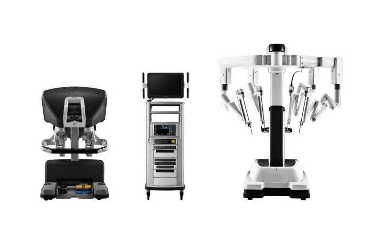
Robotic surgery in abdominal wall repair is a significant step of innovation, offering potential benefits in precision, control and safety. Leaving aside the specific issues of cost — much debated — the use of the robotic platform must be carefully weighed, considering factors such as cost, availability of adequate training and the specific needs and conditions of patients.
Beyond the specific techniques, and whatever operative mode is used in wall surgery, open or minimally invasive, the following are fundamental technical elements, the cornerstones of this surgery, which can be performed both in open surgery and with minimally invasive and robotic techniques.
What are the cornerstones of wall surgery?
What is the Rives-Stoppa repair?
The wall repair proposed by Rives and Stoppa is an important milestone in the history of hernia surgery. René Stoppa was a French surgeon specialised in general and abdominal surgery; Jean Rives was a close collaborator, and together they worked on several surgical techniques.
The technique was introduced in the 1980s. The procedure uses a large mesh placed in the preperitoneal position, behind the rectus muscles. It was originally described as an alternative for inguinal hernia repair, but was later adapted for ventral and incisional hernias.
After its introduction the technique became popular, especially in Europe, thanks to its advantages in reducing postoperative pain and its very low infection risk. The Rives-Stoppa technique is often combined with other procedures, such as the component separation technique, to treat complex hernias, and is today considered the safest way to restore a wall defect. It is performed by open, laparoscopic, endoscopic and robotic routes.
What is the TAR?
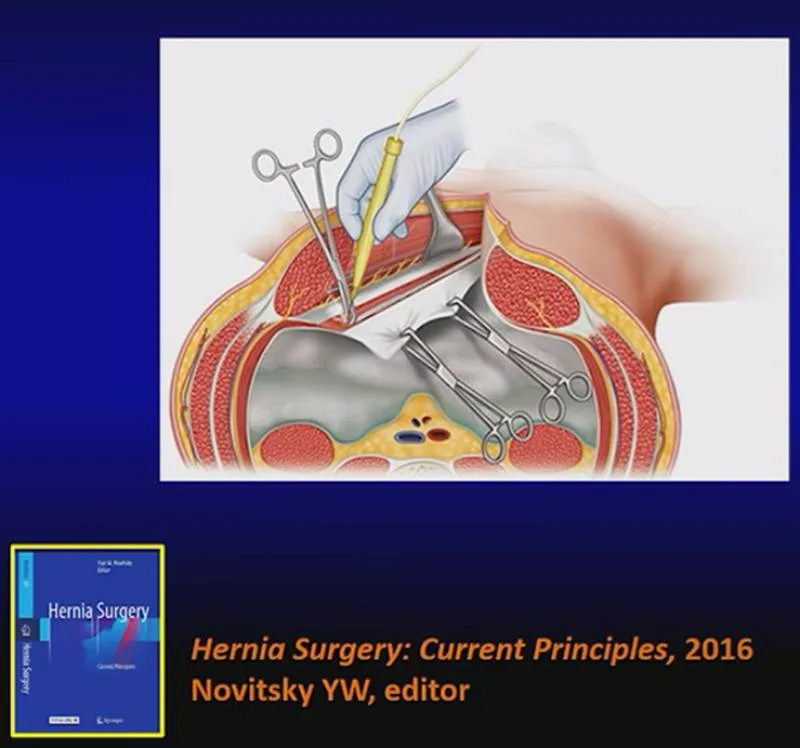
The TAR technique (Transversus Abdominis Release), proposed by Yuri Novitsky, is an advanced method used in abdominal wall repair, in particular for complex hernias. With this technique a posterior component separation (PCS) is performed.
Indications: TAR is indicated for the repair of complex abdominal wall hernias, especially with large defects, when a tension-free reconstruction of the wall is mandatory.
Technical notes: the transversus release is performed after reaching the retrorectus space. The posterior rectus sheath is incised medial to the neurovascular bundles, so as not to compromise the neurovascular supply of the rectus muscles, about 1 cm from the lateral margin of the sheath marked by the semilunar line. Beneath the posterior rectus sheath, in the upper abdomen, lies the transversus abdominis. This is incised craniocaudally (or its medial insertions are divided, as in the Madrid modification proposed by the Spanish surgeon Miguel Angel Garcia Urena). As the transversus lateralises, the dissection proceeds downward, incising the posterior sheath of the internal oblique and completing a space beneath the transversus and above the peritoneum and transversalis fascia. In this way the medial compartment of the wall is mobilised toward the midline, allowing tension-free repair of large defects. The mesh can be very wide and is placed in this same space.
TAR is an innovative and highly specialised approach for the repair of complex abdominal wall hernias. As with all surgical procedures there are potential risks and complications, and appropriate patient selection, preparation and surgical expertise are fundamental to optimal results.
When is component separation needed?
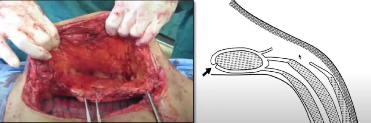
It was introduced by the plastic surgeon Dr. Ramirez in 1990 and has become one of the most commonly used techniques for the repair of complex ventral hernias, with minor modifications over time. It allows separation of the more anterior components of the abdominal wall to facilitate tension-free closure of large defects.
Indications: ACS is indicated mainly for the repair of large ventral or incisional hernias, where a tension-free closure of the wall is needed.
Technical notes: ACS involves dissecting the external oblique fascia about 1 cm from its insertion on the rectus sheath. The dissection then proceeds laterally, separating the sheath and external oblique muscle from the internal oblique. This allows wide mobilisation of the muscular components, which are medialised to repair the wall defect.
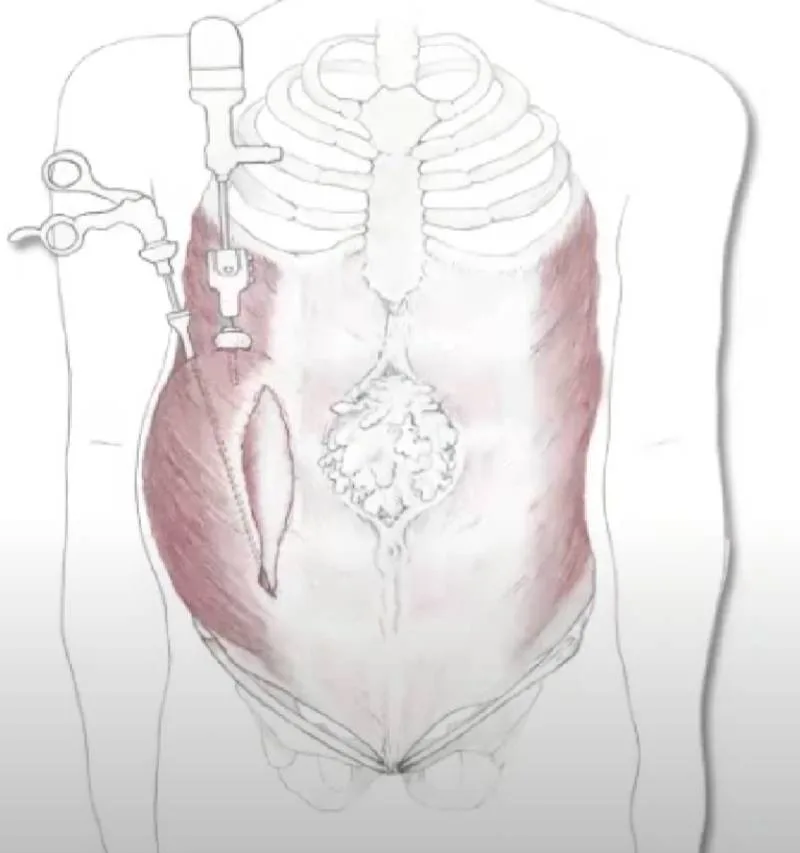
What is the endoscopic modification of component separation?
The endoscopic approach to ACS offers a minimally invasive alternative that reduces surgical trauma and potentially recovery times. The main steps are: endoscopic access, with small incisions to insert the endoscopic instruments beneath the external oblique sheath; endoscopic dissection, performed under endoscopic guidance, allowing a clear view of the anatomical planes; and component separation, performed by incising from beneath the medial margin of the external oblique sheath.
Anterior component separation is an effective method for repairing complex abdominal wall hernias, especially with large defects of the anterior fascia. The endoscopic approach is a significant evolution, offering a minimally invasive option that can reduce surgical trauma and speed healing. As with all surgical procedures, appropriate patient selection and surgical experience are fundamental to the best possible results.
What are small bites in abdominal wall closure?
The small-bites principle uses closely spaced, small sutures to close the midline, reducing tension and the risk of fascial dehiscence and incisional hernia compared with large bites.
Is the technique enough to define the result?
No. Two patients with the same defect may need two different operations, because what lies above the defect changes: skin, fat, the outcomes of previous scars. The access route and the mesh plane solve the function; the quantity and quality of the covering tissues decide the shape. The two questions are posed in the same assessment, not one after the other: if the second is deferred, the morphological result is discovered only once the operation is over.
In practice, when the morphological component is modest a minimally invasive repair is enough; when the excess skin is real it must be removed, because no retraction device produces the same effect, and the wall repair is planned together with the remodelling. The assessment method, the criteria for choice and the limits are described on the network hub: federicofiori.com.
Frequently asked questions
Which access routes exist to repair a wall defect?
Operations to repair defects of the anterior abdominal wall can be performed by an open route or by a minimally invasive route (laparoscopic, endoscopic or robotic).
What is the Rives-Stoppa technique?
The wall repair proposed by Rives and Stoppa is an important milestone in hernia surgery: it uses a large mesh placed behind the rectus muscles in the preperitoneal position and is today considered the safest way to restore a wall defect.
What is component separation?
Introduced by the plastic surgeon Dr. Ramirez in 1990, it is one of the most common techniques for complex ventral hernias and allows separation of the anterior components of the wall to facilitate tension-free closure of large defects.
What advantages does robotic wall surgery offer?
Robotics is a form of minimally invasive approach with important advantages for the patient and the surgeon in terms of precision, safety and ergonomics.