Inguinal hernia
An inguinal hernia is a protrusion of fat or bowel through a weak point of the groin above the inguinal ligament. It is common in men, does not resolve on its own, and in adults is repaired with a mesh, through one open route or one of three minimally invasive routes (TAPP, TEP, eTEP).
Key points
- An inguinal hernia is the passage of preperitoneal fat, omentum or bowel through the inguinal canal or the posterior wall of the groin, above the inguinal ligament.
- It mainly affects men: the lifetime occurrence of groin hernia is 27–43% in men and 3–6% in women (HerniaSurge 2018).
- An inguinal hernia does not close or regress on its own. In men with few symptoms, careful observation is an accepted option, but most of them end up having surgery over the following years.
- In adults, repair with a mesh is the rule. Repair by suture alone is kept for selected cases.
- There is one open route (anterior, for example Lichtenstein) and three minimally invasive routes to the same preperitoneal space (TAPP, TEP and eTEP), which can also be performed with a robot.
What it is and why it happens
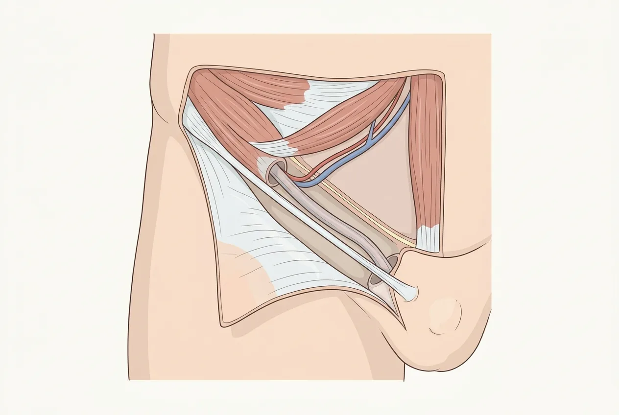
The inguinal canal is an oblique passage running from the deep inguinal ring (an opening in the transversalis fascia lateral to the inferior epigastric vessels) to the superficial ring in the external oblique aponeurosis, just above the pubic tubercle. In men it carries the spermatic cord, in women the round ligament of the uterus. Its floor is the inguinal ligament, its anterior wall the external oblique aponeurosis, and its posterior wall the transversalis fascia, reinforced medially by the conjoint tendon. The muscles that form these layers are described on the page on abdominal wall anatomy.
Seen from inside, all groin hernias leave the abdomen through the same area, which Henri Fruchaud described as the myopectineal orifice. It is bounded above by the arch of the internal oblique and transversus muscles, medially by the rectus muscle, laterally by the iliopsoas and below by the pectineal (Cooper's) ligament, and it is divided by the inguinal ligament into an upper part, where inguinal hernias form, and a lower part, the femoral canal. This is why the posterior approaches place a single mesh that covers the whole orifice: indirect, direct and femoral defects are dealt with at the same time.
The orifice is protected by a shutter mechanism. When the abdominal muscles contract, the arch of the internal oblique and transversus descends towards the inguinal ligament and narrows the posterior wall, while the deep ring is pulled upwards and laterally. A hernia forms when this mechanism fails or the tissue behind it gives way.
Indirect hernias follow the path of the spermatic cord through the deep ring. Many have a congenital origin, that is, a processus vaginalis that did not close after the descent of the testis. Direct hernias push through the posterior wall medial to the epigastric vessels and are acquired. The quality of the connective tissue plays a part in both: an altered ratio of type I to type III collagen and increased levels of matrix metalloproteinases are associated with hernia formation and recurrence, and rare connective tissue disorders such as Ehlers–Danlos syndrome carry a higher risk (HerniaSurge 2018).
The risk factors usually reported are male sex, older age, family history, a previous hernia on the other side, smoking, chronic obstructive pulmonary disease, chronic constipation, prostatism, heavy physical work and abdominal aortic aneurysm, which shares a defect of the connective tissue. Straining at stool, coughing or lifting increase pressure on a wall that is already weak. They are a trigger, not the cause: the hernia appears where the tissue was already inadequate.
Classifications, and which one is used
Many classifications of groin hernia have been proposed. An expert panel of the European Hernia Society reviewed those of Nyhus, Gilbert, Rutkow, Schumpelick, Harkins, Casten, Halverson, McVay, Lichtenstein, Bendavid, Stoppa, Alexandre and Zollinger before proposing a single system (Miserez 2007, HerniaSurge 2018). The ones that are still quoted are the following.
Nyhus
It is based on the state of the posterior wall and of the deep ring. Type I is an indirect hernia with a normal ring, type II an indirect hernia with an enlarged ring, type III a defect of the posterior wall (IIIa direct, IIIb indirect with a destroyed posterior wall, IIIc femoral) and type IV a recurrence. It was designed with the posterior approach in mind and describes what has to be reconstructed.
Gilbert
It grades the hernia during an open operation according to the size of the deep ring and the integrity of the floor, from small indirect hernias (type 1) to large direct defects (type 5). Rutkow and Robbins later added a type 6 for combined direct and indirect hernias (pantaloon) and a type 7 for femoral hernias. Its use is mostly descriptive in open surgery.
Rutkow-Robbins
It is the extended version of Gilbert described above, with the two added types. It was used to compare plug-and-patch repairs, and outside that setting it has had limited use.
Aachen (Schumpelick)
It combines the site of the hernia (lateral, medial, femoral, combined) with the diameter of the defect measured against the tip of an instrument, in three classes. Its simplicity made it the basis of the European system.
Zollinger
An updated traditional classification that keeps the distinction between indirect, direct, combined and femoral hernias and grades each by size, so that it can be applied in both open and laparoscopic surgery (Zollinger 2004).
EHS
The European Hernia Society classification, derived from Aachen, records three items: primary (P) or recurrent (R) hernia, site (L lateral, that is, indirect, M medial, that is, direct, F femoral) and size of the defect (1 up to 1.5 cm, 2 up to 3 cm, 3 over 3 cm, measured against the length of a standard laparoscopic grasper or the index finger), with an X when the size was not assessed (Miserez 2007). HerniaSurge suggests its use because it fulfils most requirements and is simple (HerniaSurge 2018). It is the classification used in operative reports and registries, because it can be applied in the same way in open and posterior repairs and it fits a single grid.
| P / R | 0 | 1 (≤1.5 cm) | 2 (≤3 cm) | 3 (>3 cm) | X |
|---|---|---|---|---|---|
| L (lateral) | |||||
| M (medial) | |||||
| F (femoral) |
| Classification | Based on | Main use |
|---|---|---|
| Nyhus | Deep ring and posterior wall, recurrence as a separate type | Planning a posterior repair |
| Gilbert | Size of the deep ring and integrity of the floor, at open surgery | Description in open surgery |
| Rutkow-Robbins | Gilbert plus combined and femoral hernias | Comparison of plug-and-patch repairs |
| Aachen (Schumpelick) | Site and diameter of the defect | Basis of the EHS system |
| Zollinger | Traditional types graded by size | Open and laparoscopic description |
| EHS | Primary or recurrent, site (L, M, F), size (1, 2, 3) | Operative reports, registries and research |
Presentation and natural history
The usual presentation is a swelling in the groin that appears on standing or straining and disappears lying down, with a dragging discomfort that grows during the day. Some hernias are found by chance at a visit for another reason. Pain that is sharp, constant and not related to the swelling suggests looking for another cause.
A hernia does not regress. In men with no or minimal symptoms, watchful waiting is an accepted option. In the North American randomised trial of 720 men, pain limiting activities at 2 years did not differ between watchful waiting and repair, 23% of the men assigned to observation had crossed over to surgery by 2 years, and acute incarceration occurred at a rate of 1.8 per 1000 patient-years (Fitzgibbons 2006). With longer follow-up the estimated crossover rate reached 68% at 10 years, 79% in men older than 65 against 62% in younger men, and the main reason was pain (Fitzgibbons 2013). Observation therefore postpones the operation in most men rather than avoiding it.
Women are managed differently. Female sex, a femoral hernia and a previous admission for groin hernia are risk factors for incarceration and strangulation (HerniaSurge 2018), and in women a groin swelling may be a femoral hernia that a clinical examination can miss. For this reason repair is generally advised rather than observation, and when expertise is available a laparo-endoscopic repair is suggested, because it shows the femoral canal and reduces the risk of chronic pain (HerniaSurge 2018).
Diagnosis
Clinical examination, standing and lying down, with a cough or Valsalva manoeuvre, is the reference and in most men is enough to plan the repair (HerniaSurge 2018). It distinguishes a reducible from an irreducible hernia and assesses the contralateral side.
Dynamic ultrasound, performed during straining, is useful when the swelling is not palpable, when pain is present without a clear bulge and to distinguish the hernia from other structures. CT and MRI are kept for uncertain cases: occult hernias, chronic groin pain in athletes, suspected complications and recurrences after several repairs, where MRI also shows the muscular and tendon insertions on the pubis. Further detail is on the page on imaging and radiology.
The differential diagnosis includes athletic pubalgia (pain at the insertion of the adductors and rectus abdominis), lipoma of the spermatic cord, hydrocele, enlarged inguinal lymph nodes, femoral hernia and, in women, endometriosis of the canal of Nuck, which causes a swelling and pain that follow the menstrual cycle.
Open repair
In open anterior repair the canal is opened through a skin incision of a few centimetres above the inguinal ligament, the hernia sac is dissected from the cord and reduced, and the posterior wall is reinforced. It can be performed under local anaesthesia, which HerniaSurge recommends in open repair provided the surgeon is experienced in the technique (HerniaSurge 2018).
Lichtenstein
Described as a tension-free hernioplasty (Lichtenstein 1989), it places a flat mesh on the posterior wall of the canal, under the external oblique aponeurosis, sutured to the inguinal ligament below and to the internal oblique above, with a slit for the cord. It is the reference open technique in guidelines and in registries. In the Danish database, recurrence at 96 months was 3% after Lichtenstein against 8% after open repair without mesh, although the authors of the guidelines note the limits of that comparison (HerniaSurge 2018).
Shouldice
A repair without mesh in which the transversalis fascia is divided and the posterior wall is rebuilt in four running layers. In high-volume expert centres it has a recurrence rate of less than 2%, but these data come mainly from those centres (HerniaSurge 2018). It remains an option when a mesh is to be avoided or is refused.
Open preperitoneal repairs (TIPP, TREPP)
They place the mesh in the preperitoneal space, the same plane as laparoscopic repair, through an open access: transinguinal in TIPP (transinguinal preperitoneal), through the rectus sheath in TREPP (transrectus sheath preperitoneal). Their aim is to obtain posterior coverage of the myopectineal orifice without pneumoperitoneum and with limited contact with the inguinal nerves.
Plug
A cone of mesh pushed into the defect, usually combined with an onlay patch (plug-and-patch). It is quick to place, but it leaves a three-dimensional implant in the canal, and its use has decreased in registries in favour of flat meshes.
Desarda
A repair without mesh that uses a strip of the external oblique aponeurosis, left attached at both ends, as a new posterior wall. It is proposed where meshes are not available or not accepted.
Open repair is the appropriate choice in several situations: when local anaesthesia is required, for large inguinoscrotal hernias, after previous pelvic surgery or radiotherapy that has obliterated the preperitoneal space, when pneumoperitoneum or general anaesthesia are contraindicated, and when no laparo-endoscopic expertise is available. The choice depends on the defect, the patient and the experience of the surgeon.
Minimally invasive repair
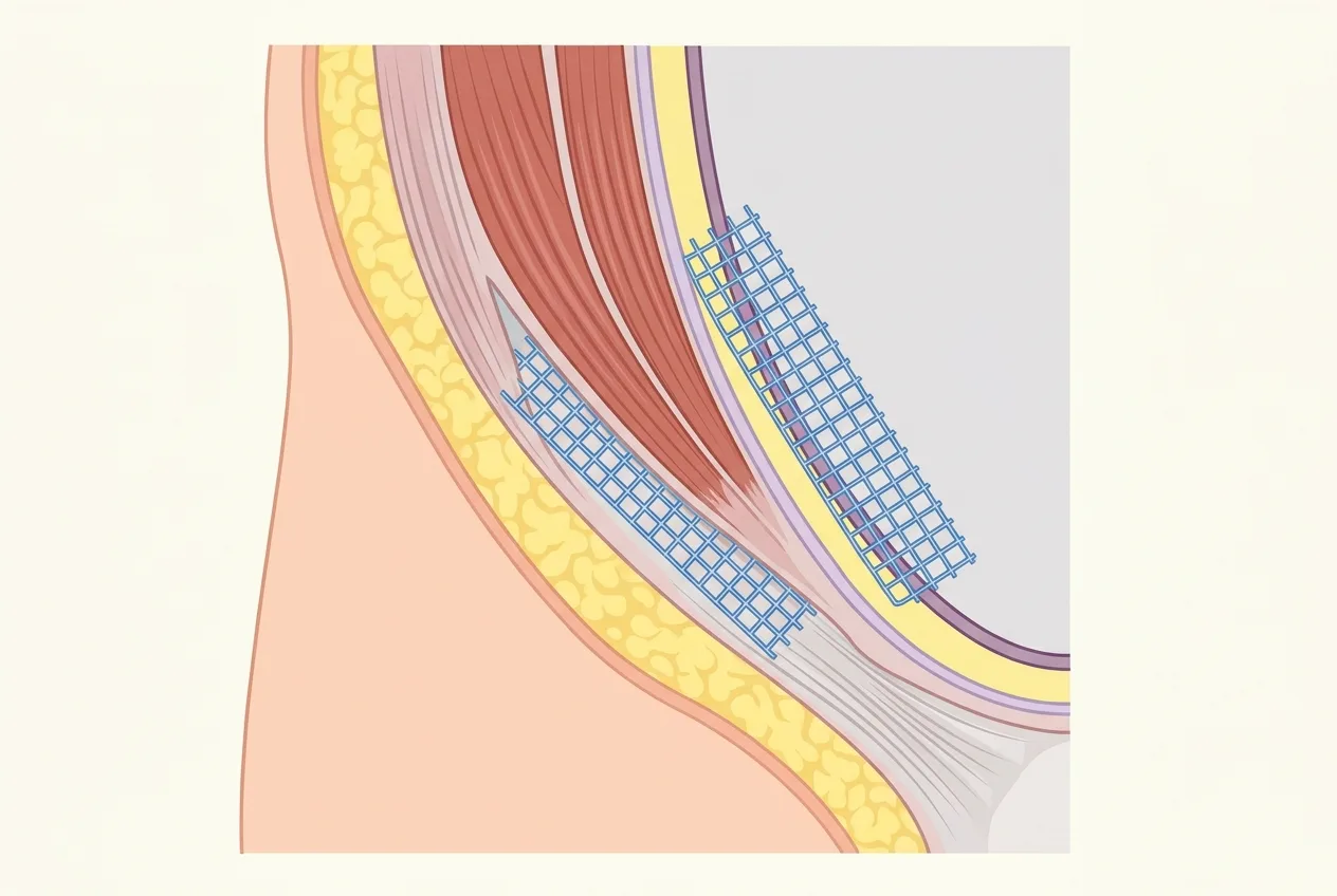
All laparo-endoscopic techniques work behind the abdominal wall, in the preperitoneal space, and cover the entire myopectineal orifice with a flat mesh. The mesh plane is the same, and what changes is how the space is reached and how the peritoneum is handled. The learning curve is longer than for Lichtenstein, especially for TEP, and ranges between 50 and 100 procedures (HerniaSurge 2018).
TAPP (transabdominal preperitoneal repair)
The abdomen is entered and both groins are seen from inside. A peritoneal flap is opened above the myopectineal orifice, the preperitoneal space is dissected, the sac is reduced, a mesh large enough to overlap the whole orifice is placed and the peritoneum is closed over it. The view of both sides and of the anatomy is wide. The risks specific to TAPP come from entering the peritoneal cavity: in the German Herniamed registry visceral injuries (bowel, bladder) occurred in 0.27% of TAPP against 0.1% of TEP, a difference that was not statistically significant (HerniaSurge 2018).
TEP (totally extraperitoneal repair)
The preperitoneal space is entered directly, below the umbilicus, behind the rectus muscle, and is developed with a balloon or with the scope, without entering the abdomen. The mesh is placed in the same position as in TAPP and there is no peritoneal flap to close. The working space is narrower, the anatomy less familiar at first, and the learning curve is the longest of the three.
eTEP (enhanced-view totally extraperitoneal repair)
Described by Jorge Daes (Daes 2011), eTEP keeps the extraperitoneal plane of TEP but changes the access: the first port is placed higher and more laterally, in the retromuscular space of the rectus, further from the defect, and the ports are arranged along a wider field. The result is a larger working space, freer movement of the instruments and an easier dissection in difficult cases such as large inguinoscrotal hernias or after radical prostatectomy, which were among the first cases treated (Daes 2011). In practice it is a TEP with more room.
TEP or TAPP
Studies comparing the two techniques show similar rates of seroma, cord and scrotal swelling, testicular atrophy, bladder injury, nerve lesions, chronic pain and recurrence (HerniaSurge 2018). In the literature analysed by the guidelines, recurrence fell between the 1990s and 2008 to 0.77% for TAPP and 0.54% for TEP (HerniaSurge 2018). The difference lies in the risk profile (intraperitoneal access for TAPP, a narrower space and more conversions for TEP) and in the ergonomics. In bilateral hernias a meta-analysis of 1,714 patients found comparable safety and efficacy, with fewer conversions to open repair in TAPP (2.92% of TEP converted against 0%) and, in a sensitivity analysis, fewer recurrences in TAPP (Oliveira Carneiro 2026). The choice also shifts for recurrence after open repair, where a posterior approach is advised because it works in untouched tissue (HerniaSurge 2018), for femoral hernias, which are covered by the same mesh, and after prostatic surgery, where the preperitoneal space may be scarred and a transabdominal view or the wider space of eTEP can help.
Robotic repair
The robot is used mainly to perform a TAPP. What it changes is the instrument: a stable three-dimensional view, wristed instruments, a seated surgeon and an easier suture of the peritoneum. It does not change the anatomy, the plane or the mesh.
In the reference randomised trial (RIVAL, 102 patients), robotic TAPP was not superior to laparoscopic TAPP in wound events, readmissions, pain or quality of life at 30 days, and it was associated with longer operative times (median 75.5 against 40.5 minutes), higher cost and greater surgeon frustration (Prabhu 2020). A later single-centre comparison by experienced surgeons reported shorter operative times with robotic TAPP, despite about 5 minutes for docking (Arunthavanathan 2025). Taken together, the data support its safety without showing a clinical benefit for the patient.
It is reasonable where the platform is already in use and the team is trained, in complex or recurrent hernias that need a long dissection, and as a training platform. In a primary unilateral hernia it is a choice of platform, not of outcome.
Mesh and fixation
Most groin meshes are made of monofilament polypropylene. Lightweight, large-pore meshes may have slight short-term advantages in pain and convalescence but are not associated with better long-term results in recurrence or chronic pain (HerniaSurge 2018). Self-gripping meshes, with microhooks on one side, were designed to reduce or avoid traumatic fixation in Lichtenstein repair (HerniaSurge 2018). Biological and biosynthetic meshes have no routine role in elective groin hernia repair and are considered only in rare contaminated settings. More on materials and planes is on the page on meshes and the plane of placement.
The mesh can be fixed with sutures (open repair), tacks, glue (cyanoacrylate or fibrin sealant) or not fixed at all, which is common in TEP for most hernias because the preperitoneal space holds it in place. No fixation method has been shown to be the best (HerniaSurge 2018). The link with pain is mechanical: every suture or tack that catches a nerve, or the periosteum of the pubis, is a possible source of chronic pain, which is why atraumatic fixation, or none, is preferred whenever stability allows it.
Complications
Seroma, that is, a collection of fluid in the space left by the sac, is common after posterior repair of large direct hernias and usually resolves on its own. Haematoma, urinary retention (more frequent in older men and after general or spinal anaesthesia) and wound infection, which is rare, are the other early complications. Meta-analyses of randomised trials have not shown different rates of urinary retention between laparoscopic and open techniques (HerniaSurge 2018). Recurrence after mesh repair by an experienced surgeon is in the order of a few per cent or less (see the figures in the sections above), and it is higher after repair without mesh.
Chronic postoperative inguinal pain (CPIP)
Chronic postoperative inguinal pain (CPIP) is defined as bothersome moderate pain that affects daily activities and lasts at least 3 months after the operation (HerniaSurge 2018). Clinically significant chronic pain occurs in 10–12% of patients and decreases over time, and debilitating pain affecting daily activities or work in 0.5–6% (HerniaSurge 2018). It is the most frequent long-term problem after groin hernia repair, more frequent than recurrence.
The mechanism can be neuropathic, from injury or entrapment of a nerve, or nociceptive, from inflammation around the mesh or a fixation point. Three nerves are involved: the ilioinguinal, the iliohypogastric and the genital branch of the genitofemoral nerve. The risk factors are young age, female sex, high preoperative pain, early severe postoperative pain, recurrent hernia and open repair (HerniaSurge 2018).
Prevention in open surgery rests on recognising the three nerves and, in selected cases, on pragmatic resection of a nerve that is in the way, while planned resection is not suggested (HerniaSurge 2018). In posterior repair it rests on avoiding fixation in the triangle of pain and the triangle of doom. When pain persists, management is suggested in a multidisciplinary team (HerniaSurge 2018): medication, nerve blocks and, when these fail, surgery that can include removal of the mesh or of a fixation point and resection of the nerves involved.
Recovery
Most repairs are performed as day surgery or with one night in hospital. Pain is greatest in the first days and decreases over the following weeks. Laparo-endoscopic techniques show clear advantages over open repair in early postoperative pain, use of painkillers and return to daily activities and work (HerniaSurge 2018), so the difference between the two approaches is felt above all in the first weeks.
Postoperative restrictions are highly variable and rarely based on evidence, and no study has shown that an early return to normal activities and work increases the risk of recurrence or complications (HerniaSurge 2018). Driving is possible when an emergency stop can be performed without pain and without the effect of painkillers that cause drowsiness. Sedentary work is usually resumed earlier than heavy work, and sport and lifting are increased progressively, guided by pain rather than by a fixed date, because the strength of the repair depends on the mesh and not on the time elapsed. A follow-up visit checks the wound, the presence of a seroma and the pain.
What the guidelines say
The international HerniaSurge guidelines of 2018 recommend a mesh-based repair for symptomatic inguinal hernia in adults and state that, provided resources and expertise are available, laparo-endoscopic techniques have faster recovery, lower risk of chronic pain and are cost-effective (HerniaSurge 2018). Their 2023 update revised ten chapters with 39 new statements and 32 recommendations, 16 of them strong, agreed with a modified Delphi method (Stabilini 2023).
The International Endohernia Society has published its own guidelines for TAPP and TEP (Bittner 2011), updated them (Bittner 2014) and endorsed the HerniaSurge guidelines (Bittner 2018). A joint project of EAES, SAGES and EHS on open versus laparo-endoscopic repair and on TEP versus TAPP is currently under way, with a public protocol on the EAES website and a literature search closed on 7 December 2025. Its conclusions are not yet available.
Who performs the operation
The operations described on this site are performed by Dr Federico Fiori, whose curriculum and publications are on the hub. The other repair options for abdominal wall defects are on the page on surgical treatment.
Frequently asked questions
What is the difference between TEP and TAPP?
Both place the same mesh in the preperitoneal space. TAPP reaches it from inside the abdomen and closes the peritoneum afterwards, while TEP reaches it from outside the peritoneum without entering the abdomen. Results are broadly similar, and the choice depends on the case and on the surgeon's experience.
Can an inguinal hernia be repaired without mesh?
Yes, with techniques such as Shouldice or Desarda, which rebuild the posterior wall with the patient's own tissue. In adults the guidelines recommend a mesh-based repair, and suture repair is kept for selected cases or when a mesh is refused.
How long does the operation take?
It depends on the technique, the size of the hernia and whether one or both sides are repaired. In the RIVAL trial the median time was about 40 minutes for laparoscopic TAPP and 75 minutes for robotic TAPP.
When can work and the gym be resumed?
Sedentary work is usually resumed within days, physical work and the gym progressively, guided by pain. No study has shown that an early return to activity increases the risk of recurrence.
Can an inguinal hernia come back?
Yes. Recurrence is uncommon after a mesh repair performed by an experienced surgeon and more frequent after repair without mesh. A recurrence after an open repair is usually treated from behind, and vice versa, so as to work in untouched tissue.
What is done if pain persists after surgery?
Pain lasting more than three months is assessed to establish whether it comes from a nerve or from the mesh. Management, ideally in a multidisciplinary team, starts with medication and nerve blocks and only then considers surgery.
Can both sides be repaired in one operation?
Yes. Posterior approaches cover both groins through the same ports, and in bilateral hernias TEP and TAPP have shown comparable safety and efficacy.
Is general anaesthesia necessary?
Laparo-endoscopic repair requires general anaesthesia. Open repair can be performed under local anaesthesia, which the guidelines recommend when the surgeon is experienced in it.
- HerniaSurge Group. International guidelines for groin hernia management. Hernia. 2018. https://doi.org/10.1007/s10029-017-1668-x.
- Stabilini C, et al. Update of the international HerniaSurge guidelines for groin hernia management. BJS Open. 2023. https://doi.org/10.1093/bjsopen/zrad080.
- Miserez M, et al. The European hernia society groin hernia classification: simple and easy to remember. Hernia. 2007. https://doi.org/10.1007/s10029-007-0198-3.
- Zollinger RM Jr. An updated traditional classification of inguinal hernias. Hernia. 2004. https://doi.org/10.1007/s10029-004-0245-2.
- Fitzgibbons RJ Jr, et al. Watchful waiting vs repair of inguinal hernia in minimally symptomatic men: a randomized clinical trial. JAMA. 2006. https://doi.org/10.1001/jama.295.3.285.
- Fitzgibbons RJ Jr, et al. Long-term results of a randomized controlled trial of a nonoperative strategy (watchful waiting) for men with minimally symptomatic inguinal hernias. Ann Surg. 2013. https://doi.org/10.1097/SLA.0b013e3182a19725.
- Lichtenstein IL, et al. The tension-free hernioplasty. Am J Surg. 1989. https://doi.org/10.1016/0002-9610(89)90526-6.
- Bittner R, et al. Guidelines for laparoscopic (TAPP) and endoscopic (TEP) treatment of inguinal hernia [International Endohernia Society (IEHS)]. Surg Endosc. 2011. https://doi.org/10.1007/s00464-011-1799-6.
- Bittner R, et al. Update of guidelines on laparoscopic (TAPP) and endoscopic (TEP) treatment of inguinal hernia (International Endohernia Society). Surg Endosc. 2014. https://doi.org/10.1007/s00464-014-3917-8.
- Bittner R, et al. Endorsement of the HerniaSurge guidelines by the International Endohernia Society. Hernia. 2018. https://doi.org/10.1007/s10029-017-1674-z.
- Daes J. The enhanced view-totally extraperitoneal technique for repair of inguinal hernia. Surg Endosc. 2011. https://doi.org/10.1007/s00464-011-1993-6.
- Prabhu AS, et al. Robotic inguinal vs transabdominal laparoscopic inguinal hernia repair: the RIVAL randomized clinical trial. JAMA Surg. 2020. https://doi.org/10.1001/jamasurg.2020.0034.
- Arunthavanathan S, et al. Shorter operative times following robotic-assisted transabdominal preperitoneal inguinal hernia repair compared to laparoscopic TAPP. Hernia. 2025. https://doi.org/10.1007/s10029-025-03402-y.
- Oliveira Carneiro R, et al. Laparoscopic totally extraperitoneal (TEP) versus transabdominal preperitoneal (TAPP) repair for bilateral inguinal hernia. Hernia. 2026. https://doi.org/10.1007/s10029-025-03515-4.